our technology platform
FLECS: The first comprehensive mechanomics platform in the world
Technology
We’re accelerating the natural evolution
of phenotypic screening:
Our FLECS platform is transforming phenotypic screening and increasing clinical relevance through a better understanding of cellular function.
Invented
1960s
1990s
Today
Method
Flow cytometry
High-content imaging
FLECS
Visual


Measures
What cells consist of
What cells look like
What cells do
Medium
Written description
Photograph
Live action
Massively Parallel
Deep dive: We visualize, image, & precisely quantify cellular contractility
Our FLECS Platform combines a vastly micropatterned elastomer that isolates contractile function in single-cells with a microscopy + computer-vision process for precisely quantifying that contractile function.
FLECS Proprietary Technology:

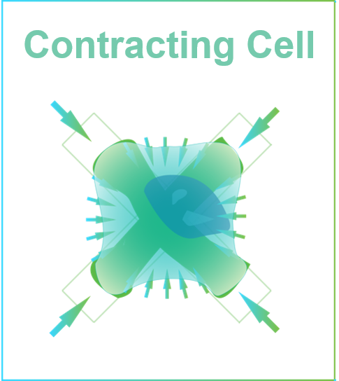
Contracting Cell
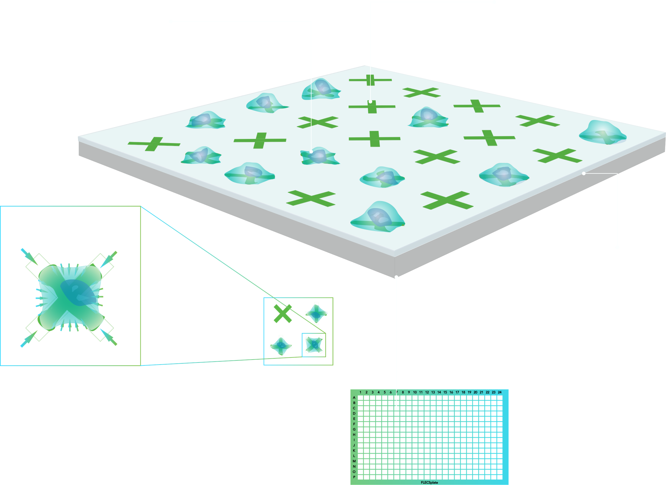
As individual cells contract, the micropatterns “shrink” proportionally to cell force. Using computer vision, micropattern changes are computed to quantify contractile force for millions of cells at a time
384-well format
Natively automation-ready. Each well provides data on 100s of single-cells
Soft, Elastomeric Layer
The well bottom is coated with transparent, planar elastomeric film that is compliant under mechanic cell stress
Adhesive Micropattern
Large, customizable arrays of fluorescent, adhesive micropatterns are embedded into the film
Adhered Single-cell
Seeded cells self-assemble over individual micropatterns, spread, and impart contractile forces
Functional. Precise. Quantitative.
Deep dive: We visualize, image, & precisely quantify cellular contractility
With FLECS, each imaged micropattern contributes a unique single-cell contractility datapoint based on how much its displaced by a bound cell. Neighboring cells are mechanically insulated from one another ensuring that each of 100s or 1000s of responding cells experience identical micro-environments and contribute unbiased data.
FLECS Proprietary Technology:
Micropattern contracted by force-generating cell
Intuitive functional cell contraction measurement you can see


Micropattern without cell
Micropattern doesn’t change unless acted on by a cell. Neighboring micropatterns are mechanically decoupled.
Fully Automated & Scalable
Platform
The FLECS platform measures contractile cell function at scale
Using lab automation and computer vision we’re able to screen drug libraries and RNAi/CRISPR libraries of any size in a cost- & time-effective manner. We’ve already run > 2M experiments – by far the most ever in a contractile function screen.
And we’re just getting started.
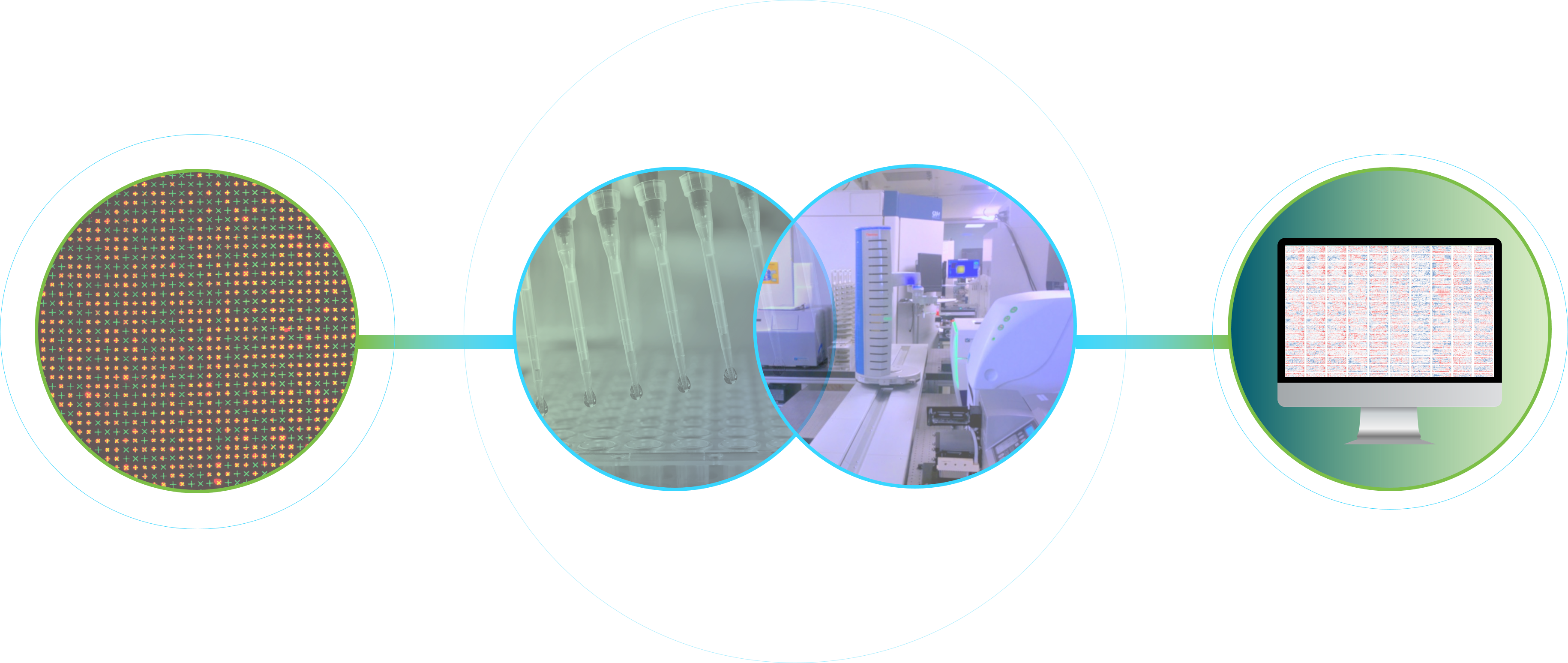
Build FLECS assay plates in-house using proprietary processes
Use powerful lab robotics to automate screening processes end-to-end
Use proprietary computer-vision to extract functional data directly linked to relevant disease pathophysiology

Integrated Platfom for Mechanomics
Solution
We’re building a new bio platform that measures mechanical cell function at scale to make better drugs, faster
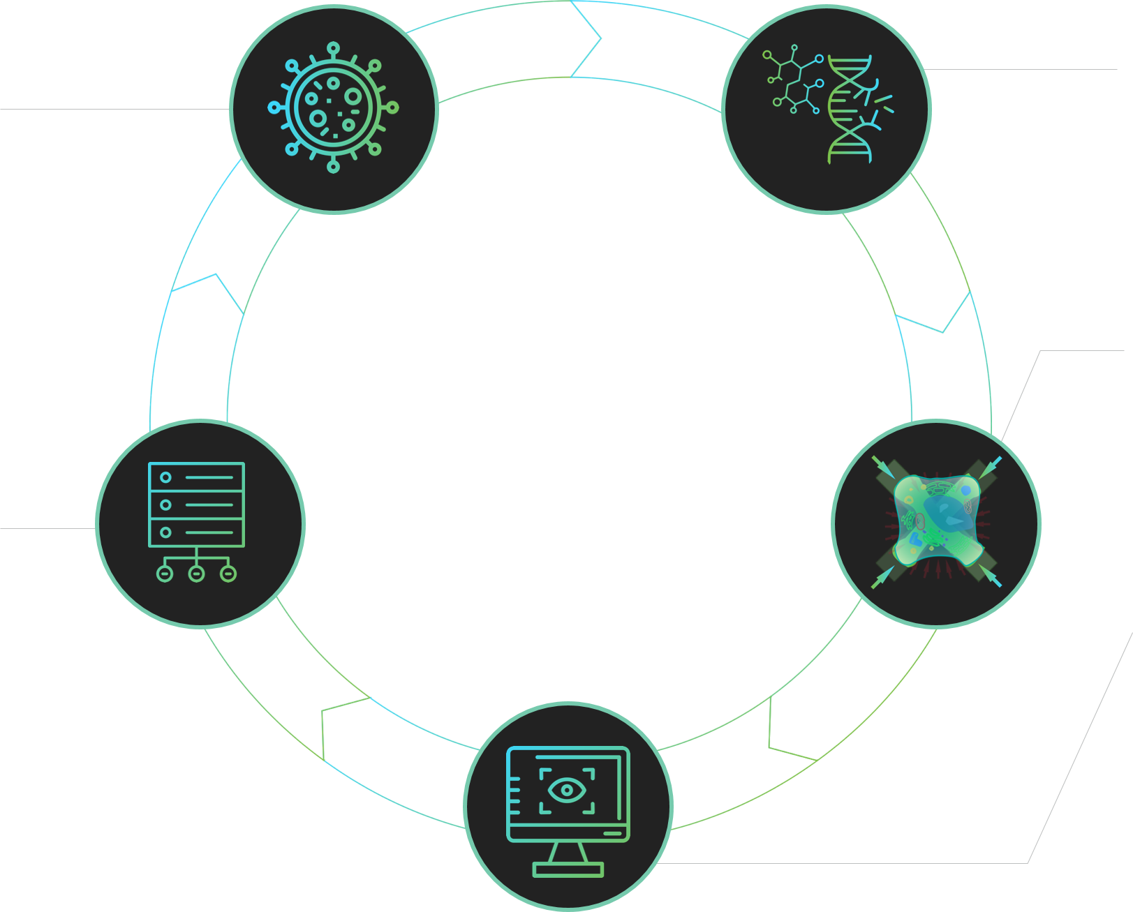
Establish Disease State
We start by establishing a diseased contractile state in primary human cells that we can screen against
Add to Our Mechanomic Database
We add the new omics scale, functional single-cell data into our growing proprietary inter-operable mechanomics data set, enabling new insights into biology

Introduce Perturbation
We perturb the cells by introducing molecules or modifying gene expression in high-throughput (up to 30k tests per week)
Visualize & Image Cell Mechano-function
Our wet-lab FLECS Platform uniformly directs cell behavior, removes inter-cell variability, and isolates mechanical function in millions of cells
Transform Images into Quantitative Data
We use computer vision to transform complex visual data in our image sets into high-res quantitative readouts of cell mechanical function at scale
